Custom Equipment
BrainSaw: automated serial-sectioning
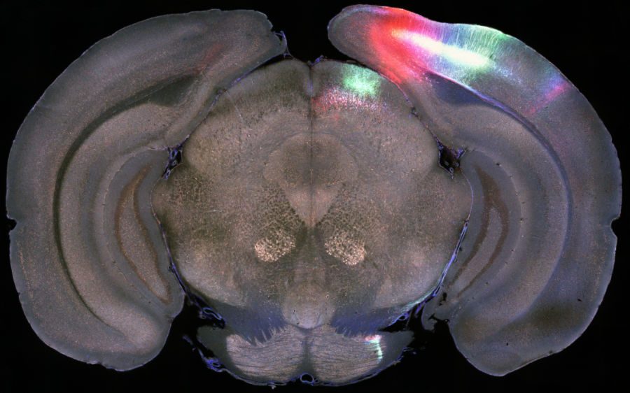
We have three ‘BrainSaw’ automated serial section microscopes. These microscopes alternate between slicing the sample and tile-scanning the exposed surface of the block. Samples simply need conventional fixation and agar embedding. The approach is generally not compatible with antibody labelling, but slices can be retrieved for further processing. Each microscope can accommodate up to 6 brains at once and imaging is usually complete in under 24 hours. Applications include mapping electrode tracks, tracing bulk projections, and automated cell counting. The systems have up to four channels and can generally handle up to three fluorophores simultaneously. Users typically run their own samples. Several training sessions are needed due to the destructive nature of the technique.
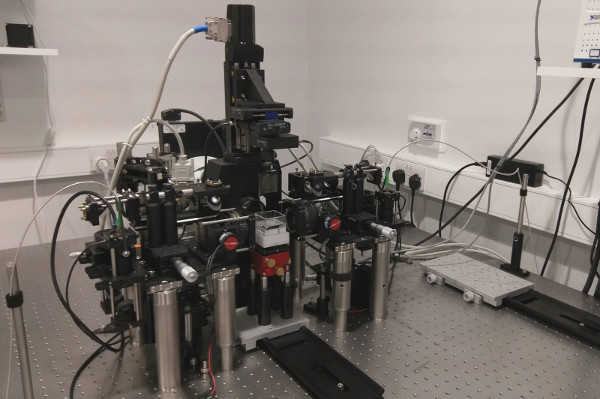
MesoSPIM: whole-brain lightsheet imaging
The MesoSPIM is a macroscope-based lightsheet microscope which provides uniform 3D resolution of cleared organs such as mouse brains and kidneys. At lower resolutions no tile scanning is required to image a mouse brain, making the microscope very fast. The system is compatible with any clearing technique. Users are responsible for clearing their own samples, but we can provide protocols and advice. The system is best suited to long term projects rather than one-off imaging sessions. Users must be trained by the AMF to have access to the microscope.
Commercial Systems
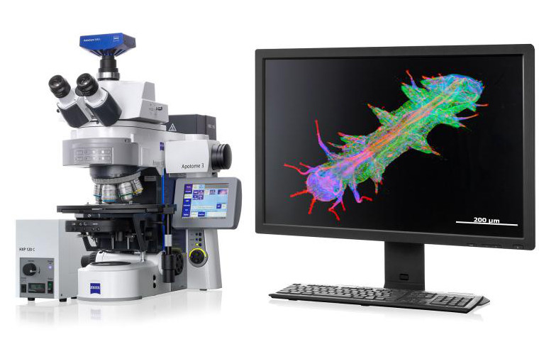
Leica SP8 Confocal
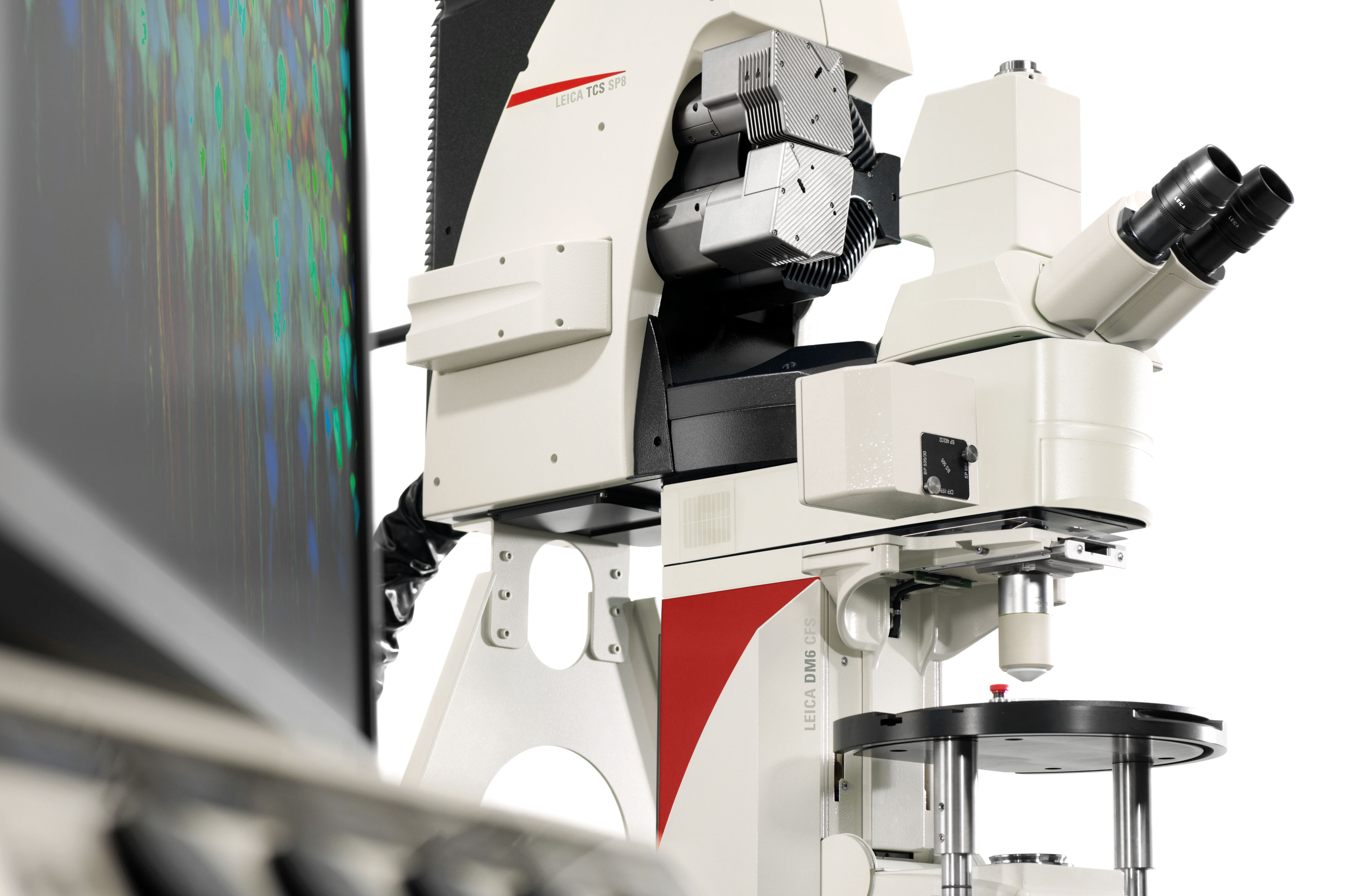
The Leica SP8 is the highest resolution microscope in the AMF. The thin optical sections produced by this confocal are ideal for imaging sub-cellular structures, such as axons and synapses. The microscope is also used for generating detailed images of cells, allowing their processes to be traced. The software has a slide-scanning feature, which allows even thick cleared slices to be imaged with ease. A white light laser with tunable excitation filter and three detectors (2x HyD and 1x PMT) with tunable emission filters provide enormous flexibility. The microscope is located in the AMF and you must be trained to have access to it.
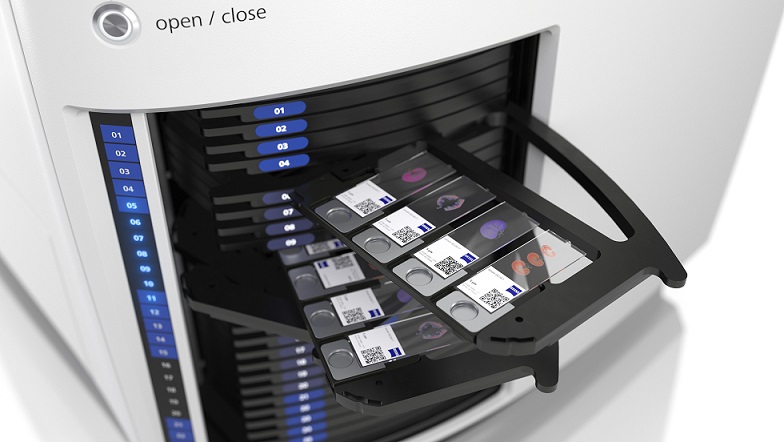
Axioscan slide scanner
This high-throughput widefield microscope accommodates up to 100 slides, which it images automatically after an interactive setup process. The system handles both fluorescence and conventional brightfield. We run this microscope alongside our histology service, but it can also be booked and used independently. You must be trained to have access to this microscope.
Widefield microscopes
We have two Zeiss Imagers, equipped with Apotomes for optical sectioning. These microscopes are ideal for imaging small numbers of slides interactively and have tile scanning ability. We also have one Zeiss Axiozoom for imaging larger fields of view. You can use this microscope for checking injection locations in whole brains, taking images of culture plates, etc. These microscopes are located on the first floor. Please contact the AMF for an induction if you are unfamiliar with them.
Functional Imaging
The AMF helps manage in vivo multi-photon microscopes across the building. Among other equipment, we manage a Thorlabs MesoScope, three 3-photon microscopes, and a Mini2p.
Available equipment at UCL
Our researchers also have access to equipment elsewhere at UCL, in particular we have links with the UCL Biosciences Imaging Facility. Equipment there includes spinning disk, high content screening, micro CT, and super resolution (SIM, PALM, STORM).